10/10/2019
Khuzema Darugar,
CADTech USA LLC
Microtia is a congenital deformity of the outer ear in children where the pinna is underdeveloped during birth. According to statistics, microtia occurs in the range of 1 to 5 out of every 10,000 births.1 In many cases, surgery is used to reconstruct the external ear of the child. Before a reconstruction surgery takes place, a surgeon performs a thorough examination to decide how to reconstruct the ear. Currently there are two ways to reconstruct the ear; using the cartilage or skin of the patient themselves, or using a prosthetic. In this article we will focus on the latter method and how 3D scanning can help simplify the process of creating a prosthetic for reconstructing an ear and how it can yield a more realistic and symmetrical ear.
3D Scan of right ear
Mirror copy of left ear
Before 3D scanning was available, surgeons carved the ear out of the patient’s cartilage from their rib cage. This can be a painful method for the patient and may lead to other complications down the road. With 3D scanning (a non-contact method) the ear is easily scanned in high resolution and then 3D printed. 3D scanning allows the surgeon to capture the intricate geometry of the human ear. In addition, it allows the capturing of the entrance to the ear canal, and the area between the ear and the head. Having a realistic replica of the patient’s ear and the surrounding head is invaluable in reconstructing a more natural looking ear.
The process of 3D scanning an ear is completely non-intrusive and takes only a few minutes. During the procedure the patient is seated on a stool, and their ear is scanned using a handheld 3D scanner (in our case, the Artec Space Spider). The 3D scanner is used to capture the outer ear, the entrance of the ear canal, and the space between the back of the ear and the head. Once the scanning is completed, the scans are aligned, cleaned and texturized in the Artec Studio software.
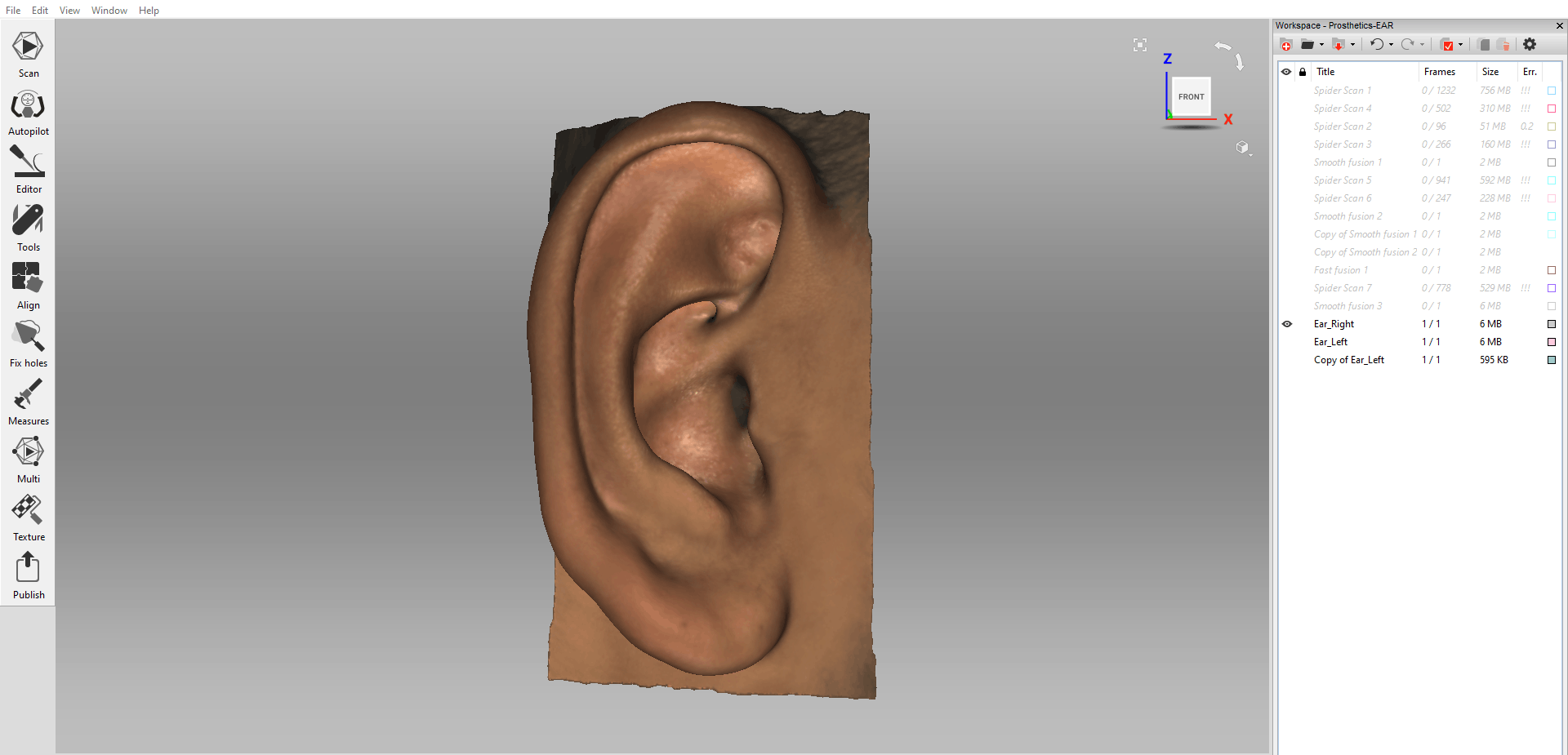
Scanned right ear shown in Artec Studio
Once the scanning is completed of the initial ear, a mirror copy is created for the opposite deformed ear. The mirror copy of the scanned ear will be used by the surgeon to create the prosthetic for the deformed ear, and also used as visual aid in preparation for the surgery. At times, a scan of the deformed ear is also taken for use as a reference.
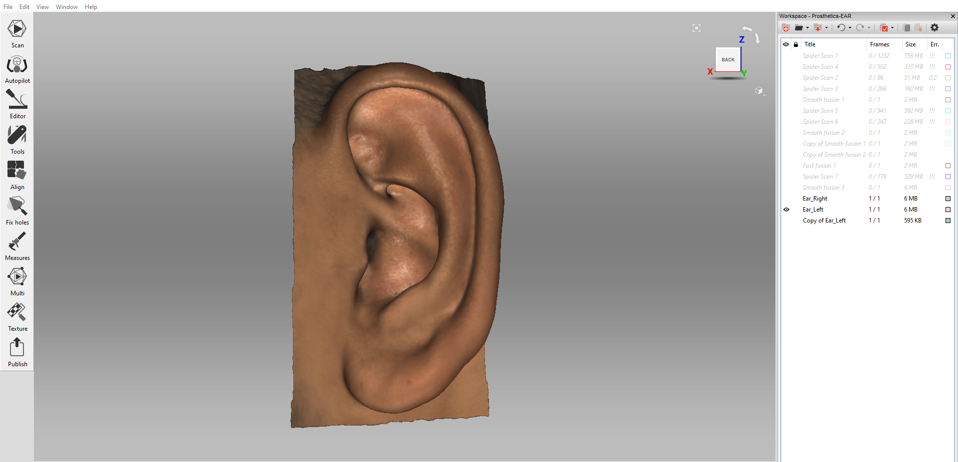
Mirrored left ear shown in Artec Studio
After creating the digital replicas of both ears, the scanned mesh of both ears is exported as STLs and sent to the surgeon for further processing. The surgeon will usually create an enlarged sized copy of the ear to the predicted adult size of the ear of the child. The copy is then sent for 3D printing using specialized materials.
In summary, 3D scanning in combination with 3D printing is the future in ear reconstruction surgery. The advantages 3D scanning brings to the table will fundamentally change how new ears are reconstructed while simplifying the process of capturing the precise geometry of an ear.
References:
1. Facts about Anotia/Microtia | CDC. (2018, November 1). Retrieved from https://www.cdc.gov/ncbddd/birthdefects/anotia-microtia.html.


